Clinical Case Example
Reconstruction of the mandible using a 3D printed titanium implant with mesh scaffold
Challenge
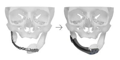
A male patient was diagnosed with a benign tumor in the medial part of the mandible. The patient underwent radical resection of the tumor with a surgical margin and immediate mandibular reconstruction with a standard plate. Due to complications after this first reconstruction, the patient required a revision surgery.
The main goal was to design a suitable implant, with adequate mass, which would ensure good bone integration and restore the appearance of the original mandible.
Solution
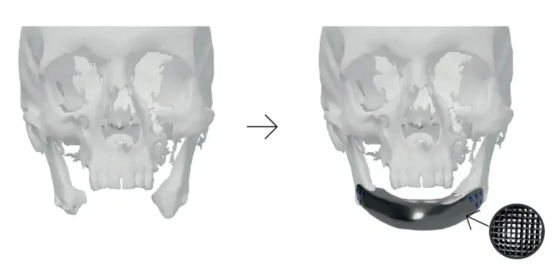
During the 3D preoperative planning, the optimal mandibular position was defined by the virtual repositioning of the mandible. The next step was to design a patient-specific implant to "bridge the gap" according to the surgeon’s guidance.
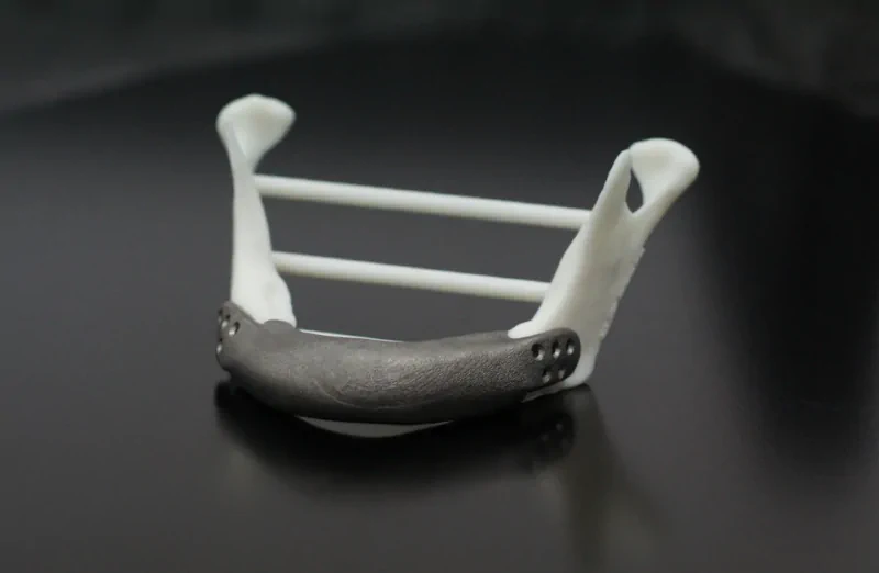
Because of the patient’s previous history and to ensure optimal bone integration, we proposed the design of the implant with an internal mesh structure. Such a porous structure with porosity 60 – 80% and appropriate pore sizes is optimal for maximal osseointegration. By using a porous structure full integration between bone and mesh is possible, and what is more, the implant itself loses weight.
The surgical guide was designed after deterrmining the location and shape of the implant in such a way that it matched the patient’s anatomy perfectly. This drilling guide indicated the outline of the planned mandible reposition and allowed to prepare mandible for the implant fixation.
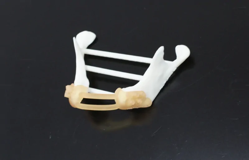
When the planning and the design were accepted by the surgeon, we started manufacturing the Customy RecoBox, which contained the patient-specific implant, surgical guide, and anatomical models. The porous titanium alloy implant was manufactured using 3D technology (electron-beam melting). The surgical drilling guide was 3D printed from a sterilizable material that can be used for short-term in a patient. The RecoBox contained also anatomical models of the patient’s mandible before and after reconstruction which helped the surgeon to tangible visualize the patient’s problem and to practice his surgical approach before the surgery.
Surgery
During the surgery, resection of the previous plate was performed and bone surfaces were prepared for the new patient-specific titanium implant. The surgical drilling guide was used for making precise holes for the implant fixation. 10 CFM titanium screws were used to fix the implant for preliminary fixation. Full fixation should be achieved after bone osseointegration. As the last stage of this procedure, soft tissues were reconstructed.
Results
The preoperative surgery plan was transferred to the OR by using the surgical drilling guide. This designed surgical guide fitted exactly on a predetermined part of the mandible and allowed to drill holes for implant fixation at the right place and angle. Thanks to that preparation, the implant placement and fixation went smoothly. There were no complications with the implant after the surgery. The results of the surgery were more than satisfactory.