


In today’s world, it is crucial to eliminate sensitive information from medical data. Dicom Anonymization is an effortless way to keep your patient and his study private. With less than few steps, the DICOM image series can be cleared from its labels.

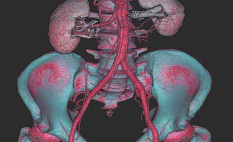
A way from CT scan to mesh model starts with highlighting anatomical structures. Basic Segmentation is a tool designed to obtain segmentation masks. By using thresholding, automatic histogram analysis or Point&Grow methods, the user can create one, that fits his needs. As a result, anatomical structures can be easily distinguished from each other.

There are cases, where volume quality is far from acceptable or there is no other option to get it good. Artefacts, noises, blurs – all of that can be reduced by Volume Filtering. Thanks to this tool, even bad quality volume may be used. That dismisses the necessity to acquire a new volume and expose the patient to another dose of radiation.

Segmentation mask filtering may come in handy when the created mask has some errors or noises caused by artefacts registered in the CT study. The mask can be morphed, smoothed, denoised or undergo Boolean operations with another mask.

A mesh model is often a raw representation. With the use of Surface Modelling, their structure is edited, modified and repaired. When needed, models can be paired together or subtracted, even their common parts can be shown. All this changes a blocky mesh to a post-processed model, which is an anatomical representation.

For ongoing therapies, where for example cancer growth must be controlled, it is recommended to use Volume Registration. There, the user combines two volumes and match them for the best possible fit. In the end, it is easier to spot a difference in size obtained over time, especially by exporting a mask from one volume to another.


